Department Research Areas
Cardiovascular ENgineering
Research in cardiovascular engineering focuses on understanding the mechanisms, treatments, and detection of cardiovascular disease. This includes 1) applying fluid dynamics and solid mechanics concepts to study blood flow, thrombosis, and vascular remodeling, 2) studying medical devices and therapeutics used to treat cardiovascular disease, and 3) using medical imaging to study cardiovascular disease progression in animal models.
Participating Faculty
The following faculty work on research around cardiovascular engineering. Visit their profile pages to learn more about their specific areas of research.
Sample Projects
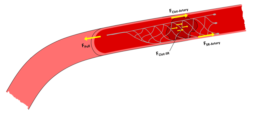
Modeling of mechanical thrombectomy for stroke
Acute ischemic stroke (AIS), the result of embolic occlusion of a cerebral artery, is responsible for 87% of the 6.5 million stroke-deaths each year. Despite improvements in stent-retriever and aspiration devices, 80% of eligible AIS patients will either die or suffer a major disability. In order to improve patient outcomes, the underlying biomechanics governing the removal of blood clots from cerebral arteries need to be better understood and improved surgical therapies developed. Dr. Good’s Cardiovascular Biomechanics Lab is utilizing both experimental and computational models of AIS to investigate the effects of patient-specific geometries, blood clot properties, and hemodynamic conditions on successful clot removal.
Representative publications:
Froehler M, Good B. The fluid mechanics of aspiration thrombectomy. Journal of NeuroInterventional Surgery. 2025.17:759-763. https://doi.org/10.1136/jnis-2024-022780
Poulos D, Froehler M, Good B. Investigation of stent retriever removal forces in an experimental model of acute ischemic stroke. Frontiers in Neurology. Vol 15. 2024. https://doi.org/10.3389/fneur.2024.1486738
Poulos D, Keith J, Froehler M, Good B. Experimental evaluation of the plunger technique: A method of cyclic manual aspiration thrombectomy for treatment of acute ischemic stroke. Interventional Neuroradiology. 2024. https://doi.org/10.1177/1591019924123036
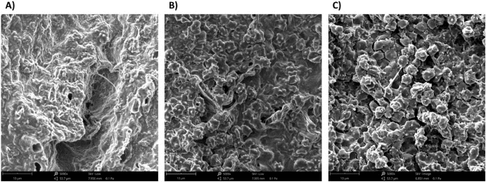
Mechanical and molecular properties of blood clots
Venous thromboembolism and ischemic stroke affect over 1.5 million people yearly. Both diseases are a result of blood clots forming in venous or arterial vasculature. Characterizing the spatial heterogeneity (Crouch Lab) and mechanical properties (Good Lab) of blood clots is important for understanding disease initiation/progression. Molecular spatial heterogeneity can contribute to variations in mechanical properties, and we are currently using MALDI imaging to assess this spatial heterogeneity.
Representative publications:
McDonald RG, Poulos DA, Woodall B, Gutzwiller L, Sheth RA, Good BC, Crouch AC. A MALDI Mass Spectrometry Imaging Sample Preparation Method for Venous Thrombosis with Initial Lipid Characterization of Lab-Made and Murine Clots. Journal of the American Society for Mass Spectrometry. 2023 Jul 13;34(9):1879-89.
Good B. The influence of blood composition and loading frequency on the behavior of embolus analogs. Journal of the Mechanical Behavior of Biomedical Materials. 2023 Apr 1;140:105738.
Cardio-oncology
Despite the efficacy of chemotherapeutic agents such as doxorubicin in targeting and eliminating neoplastic growth, cardiotoxicity remains a significant problem in oncologic and cardiovascular research. Traditional 2D echocardiography currently remains the gold standard for assessing left ventricular ejection fraction (LVEF), the most widely used clinical metric for characterizing systolic functioning. This imaging modality lacks the sensitivity needed for detecting regional myocardial dysfunction, rendering it incapable of identifying subclinical cardiotoxicity. Numerous studies have shown global longitudinal strain to be an independent predictor and unrivaled metric for prognosis in patients with reduced and preserved LVEF. To address the limitations of the ejection fraction metric, we use a novel 4D ultrasound (4D US) strain-imaging for early detection of myocardial changes during chemotherapy treatment. Additionally, the mechanism of drug-induced toxicity is unclear. Mass spectrometry imaging can be used to study the biomolecular changes occurring in the vasculature and heart tissue.
Webinar: HTXnext episode discussing preliminary results
Representative publication:
Fox AG, Buonodono KE, Jones AR, Thomas M, Scheper LE, Earl CC, Goergen CJ, Crouch AC. Multimodal Study of Murine Cardiovascular Remodeling: Four-Dimensional Ultrasound and Mass Spectrometry Imaging Journal of Visualized Experiments. 2025 Jan 10; doi: 10.3791/67347